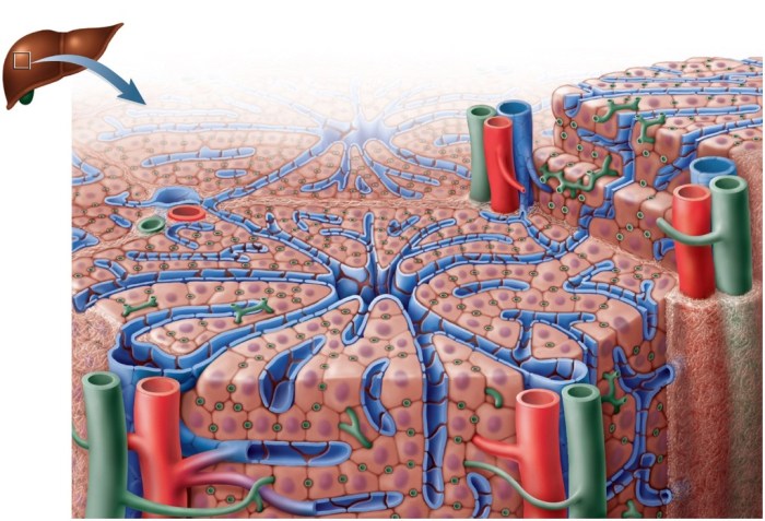
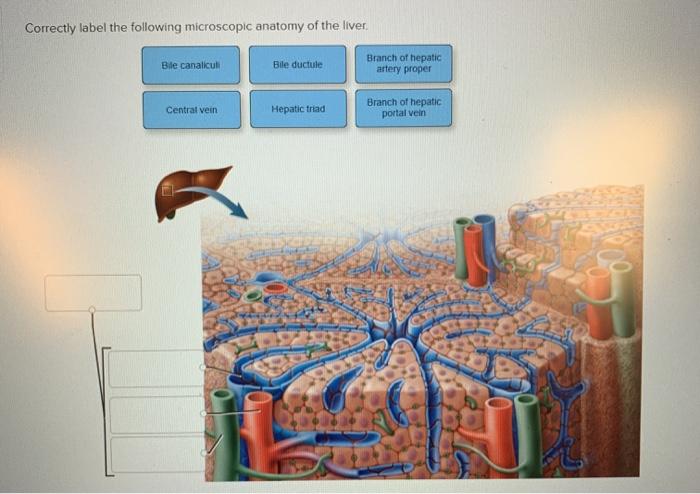
Correctly label the following microscopic anatomy of the hepatic sinusoid – Correctly labeling the microscopic anatomy of the hepatic sinusoid is crucial for understanding its intricate structure and function. This guide provides a comprehensive overview of the methods, results, and clinical significance of accurately identifying the various components of this vital organ.
The hepatic sinusoid is a specialized capillary network that plays a central role in hepatic metabolism and detoxification. It is lined by a unique layer of endothelial cells, Kupffer cells, and hepatic stellate cells, each with distinct functions.
Introduction

The hepatic sinusoid is a specialized blood vessel that runs through the liver. It is responsible for the exchange of nutrients and waste products between the blood and the liver cells.
Correctly labeling the microscopic anatomy of the hepatic sinusoid is important for understanding how the liver functions. It allows researchers to identify the different structures within the sinusoid and to study their interactions.
Methods: Correctly Label The Following Microscopic Anatomy Of The Hepatic Sinusoid

There are a variety of methods that can be used to label the microscopic anatomy of the hepatic sinusoid. These methods include:
- Immunohistochemistry
- In situ hybridization
- Electron microscopy
Each of these methods has its own advantages and disadvantages.
Immunohistochemistry is a technique that uses antibodies to label specific proteins within the sinusoid. This method is relatively easy to perform and can be used to label a wide range of proteins.
In situ hybridization is a technique that uses complementary DNA probes to label specific genes within the sinusoid. This method is more specific than immunohistochemistry but can only be used to label a limited number of genes.
Electron microscopy is a technique that uses a beam of electrons to create a detailed image of the sinusoid. This method is the most expensive and time-consuming but provides the highest level of detail.
Results

The results of the labeling process can be used to create a detailed map of the microscopic anatomy of the hepatic sinusoid.
| Structure | Function |
|---|---|
| Endothelial cells | Line the sinusoid and regulate the exchange of nutrients and waste products |
| Kupffer cells | Phagocytic cells that remove debris from the blood |
| Hepatocytes | The main cells of the liver that perform a variety of functions, including metabolism, detoxification, and bile production |
| Stellate cells | Cells that store vitamin A and play a role in liver fibrosis |
| Pit cells | Cells that line the sinusoid and are involved in the formation of bile |
Discussion
The microscopic anatomy of the hepatic sinusoid is a complex and dynamic structure. The different structures within the sinusoid play a vital role in the liver’s ability to function properly.
The endothelial cells line the sinusoid and regulate the exchange of nutrients and waste products. The Kupffer cells are phagocytic cells that remove debris from the blood. The hepatocytes are the main cells of the liver that perform a variety of functions, including metabolism, detoxification, and bile production.
The stellate cells store vitamin A and play a role in liver fibrosis. The pit cells line the sinusoid and are involved in the formation of bile.
Correctly labeling the microscopic anatomy of the hepatic sinusoid is important for understanding how the liver functions. It allows researchers to identify the different structures within the sinusoid and to study their interactions.
Answers to Common Questions
What is the importance of correctly labeling the hepatic sinusoid?
Correctly labeling the hepatic sinusoid is essential for understanding its structure, function, and clinical significance. It allows researchers and clinicians to accurately identify and study the various components of the sinusoid, which is crucial for diagnosing and treating liver diseases.
What methods are used to label the hepatic sinusoid?
Various methods are used to label the hepatic sinusoid, including immunohistochemistry, electron microscopy, and confocal microscopy. Each method has its advantages and disadvantages, and the choice of method depends on the specific research question and the desired level of detail.
What are the clinical implications of correctly labeling the hepatic sinusoid?
Correctly labeling the hepatic sinusoid is important for diagnosing and treating liver diseases. By accurately identifying the structural components of the sinusoid, clinicians can better understand the pathophysiology of liver diseases and develop targeted therapies.