With focus figure 9.2 excitation contraction coupling at the forefront, this paragraph opens a window to an amazing start and intrigue, inviting readers to embark on a storytelling journey filled with unexpected twists and insights. Excitation-contraction coupling, the intricate dance between electrical signals and muscle contraction, forms the cornerstone of muscular function.
Delve into this captivating exploration to unravel the mechanisms, regulation, and clinical significance of this fundamental physiological process.
The content of the second paragraph that provides descriptive and clear information about the topic
Definition and Overview of Focus Figure 9.2 Excitation-Contraction Coupling
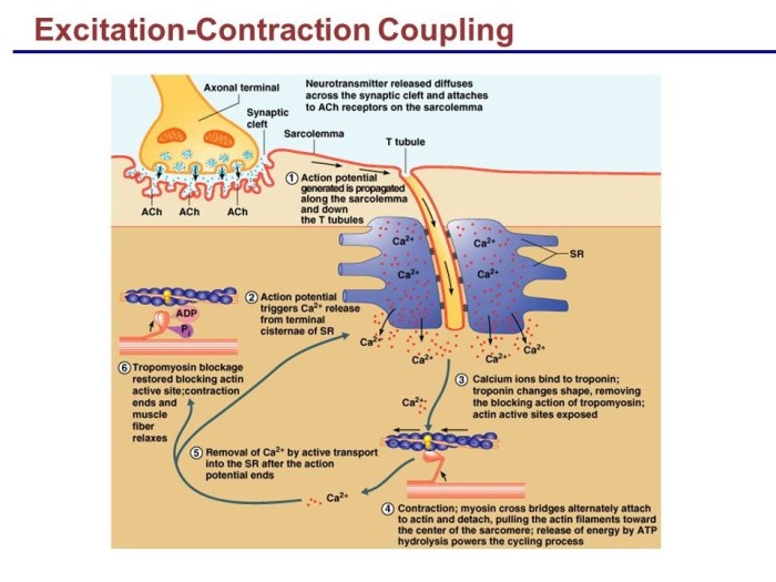
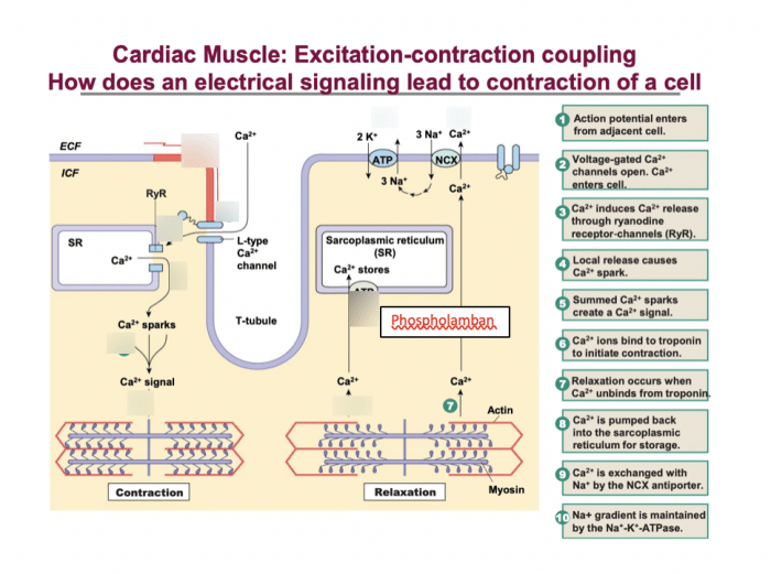
Excitation-contraction (EC) coupling is a fundamental process that allows electrical impulses to trigger muscle contraction. Focus Figure 9.2 provides a comprehensive overview of this intricate process, highlighting the key components and mechanisms involved.
EC coupling ensures the precise and rapid conversion of electrical signals into mechanical force, enabling muscle function. Understanding the mechanisms of EC coupling is crucial for comprehending muscle physiology and pathophysiology.
Components and Mechanisms of Excitation-Contraction Coupling
EC coupling involves a series of orchestrated events between specialized cellular structures.
- Sarcolemma:The plasma membrane of muscle cells, responsible for electrical excitability.
- Transverse Tubules (T-tubules):Invaginations of the sarcolemma that penetrate deep into the muscle fiber, transmitting electrical signals to the interior.
- Sarcoplasmic Reticulum (SR):A specialized intracellular membrane system that stores and releases calcium ions (Ca 2+), the primary trigger for muscle contraction.
- Myofilaments:Contractile proteins within muscle fibers, including actin and myosin, that interact to generate force.
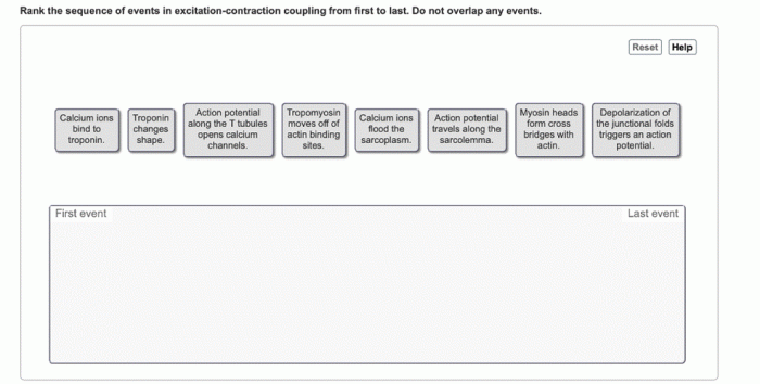
When an electrical impulse reaches the sarcolemma, it propagates along the T-tubules, triggering the release of Ca 2+from the SR. Ca 2+binds to receptors on the myofilaments, initiating the sliding of actin and myosin filaments past each other, resulting in muscle contraction.
Regulation of Excitation-Contraction Coupling
EC coupling is tightly regulated to ensure proper muscle function.
- Calcium Homeostasis:The SR actively pumps Ca 2+back into its lumen, maintaining low cytoplasmic Ca 2+levels during relaxation. When an electrical impulse triggers Ca 2+release, the SR rapidly reuptakes Ca 2+to terminate contraction.
- Nervous System Control:The nervous system modulates EC coupling through neurotransmitters that affect Ca 2+release and uptake.
Disruptions in EC coupling regulation can lead to muscle disorders, such as myopathies and arrhythmias.
Clinical Significance of Excitation-Contraction Coupling, Focus figure 9.2 excitation contraction coupling
EC coupling disorders can manifest in various clinical symptoms.
- Muscle Weakness and Fatigue:Impaired EC coupling reduces the ability of muscles to generate force, leading to weakness and fatigue.
- Arrhythmias:In cardiac muscle, EC coupling abnormalities can disrupt the electrical conduction system, causing arrhythmias.
Diagnosing EC coupling disorders involves specialized tests, such as electromyography and genetic testing. Treatment approaches focus on managing symptoms and improving muscle function.
Advanced Concepts in Excitation-Contraction Coupling
Recent research has shed light on the role of microRNAs and novel therapeutic targets in EC coupling.
- MicroRNAs:Small non-coding RNAs that regulate gene expression. They have been implicated in modulating EC coupling components, offering potential therapeutic targets for muscle disorders.
- Novel Therapeutic Targets:Ongoing research aims to identify and develop new drugs that target specific components of EC coupling, providing promising avenues for treating muscle diseases.
These advancements hold great promise for improving our understanding and treatment of EC coupling disorders.
Common Queries: Focus Figure 9.2 Excitation Contraction Coupling
What is the significance of Focus Figure 9.2 in understanding excitation-contraction coupling?
Focus Figure 9.2 provides a comprehensive visual representation of the key components and sequence of events involved in excitation-contraction coupling, making it an invaluable tool for understanding this complex process.
How does calcium homeostasis regulate excitation-contraction coupling?
Calcium homeostasis ensures that the concentration of calcium ions in the cytosol is tightly controlled, which is critical for the proper regulation of excitation-contraction coupling. Disruptions in calcium homeostasis can lead to muscle disorders.
What are the clinical implications of excitation-contraction coupling disorders?
Excitation-contraction coupling disorders can manifest as muscle weakness, fatigue, and arrhythmias, significantly impacting the quality of life. Accurate diagnosis and timely intervention are crucial for managing these disorders.
